

| Ward | Maternity Ward | D.O.B/Age | 27 y.o |
| Consultant | Dr C. Hudson |
Abnormal result
APT test positive.
Presenting complaint
?haemolytic disease of the newborn
History
27 y.o. female
G2P1 at 34 weeks
RH negative with Rhesus iso-immunization. Anti-D titres 1:128. (Blood group AB negative). Coombs test positive. Risk of haemolytic disease of the new-born.
Examination
Not applicable/difficult to examine foetus in-utero. Ultrasound of the middle cerebral artery peak systolic velocity was suggestive that the baby was anaemic.
Laboratory investigations
U/s guided chordocentesis done and foetal blood sample obtained in utero. FBC whilst in utero showed Hb: 9.8.
Clinician requested APT test to ensure that foetal sample obtained during chordocentesis.
Haematology did Kleihauer Betke Test and it showed 100% foetal haemoglobin. APT test also correlated and showed foetal haemoglobin.
Other investigations
Ultrasound: middle cerebral artery peak systolic velocity suggestive that the baby was anaemic. U/s guided chordocentesis done and foetal blood sample obtained in utero. FBC whilst in utero showed Hb: 9.8.
Final diagnosis
APT test confirmed that foetal blood had been obtained during chordocentesis. It also correlated with the Kleihauer Betke done by haematology.
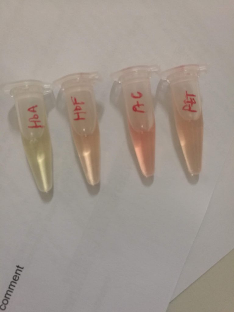
HbA: adult haemoglobin; HbF: foetal haemoglobin; PtC: patient control; PtT: test control.
Note the slight green tinge of HbA
Take-home messages
Principles of the APT test: Sodium hydroxide (NaOH) denatures adult oxyhaemoglobin to haematin (with a colour change from pink to yellow green). Foetal haemoglobin resists alkaline denaturation by NaOH and maintains a pink colour. If adult haemoglobin (HbA) is present in the sample, it turns yellow and then green within two minutes of the addition of sodium hydroxide. Any pink colour that persists for longer than 2 minutes indicates foetal haemoglobin (HbF) is present in the sample.
The Kleihauer Betke Test is an acid-elution assay performed on maternal blood to determine the amount of HbF that has passed into maternal circulation. The process exposes maternal blood smear to an acid solution. HbF, being resistant to the acid, removes intact, whereas HbA is removed. Following this, the smear is stained via Shepard’s method. The foetal red blood cells are left rose-pink in colour, and the maternal cells appear “ghost-like” due to the absence of staining. This test is done by Haematology. Other ways of differentiating maternal from foetal blood is examining the MCV. The foetus has a larger MCV. This is nonspecific though as the mother may have macrocytic anaemia.


